Radiology Corner: Case #8
The latest Radiology Corner is presented by Dr Thomas Bate, and investigates a patient experiencing an unusual presentation of bilateral hip and lumbosacral pain.
- A 75-year-old male patient presented to the pain clinic with bilateral hip and lumbosacral pain. At the same time, he presented to the colorectal surgeons reporting rectal bleeding and a change in his bowel habit. A pulsatile mass within the pelvis could be felt on digital rectal examination.
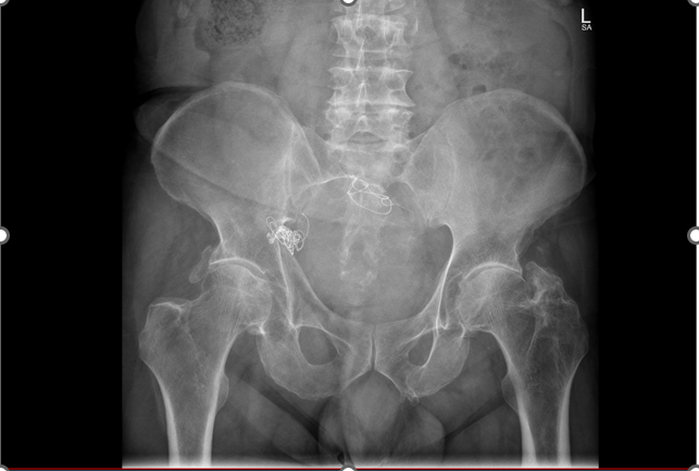
- Relevant past medical history: This patient was involved in a road traffic accident 25 years previously requiring significant vascular and bony reconstruction. Large amount of metalwork were noted on plain x-ray of the pelvis. The hip joints showed moderate arthritis on plain x-ray, more so on the left than on the right.
-
View the x-ray below. Apart from moderate osteoarthritis, what else do you note?
Click Below for Answers
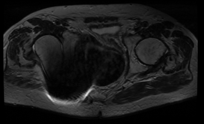
An MRI scan of his pelvis was requested, see images below. What do you notice?
Click below for answers.
Further Steps:
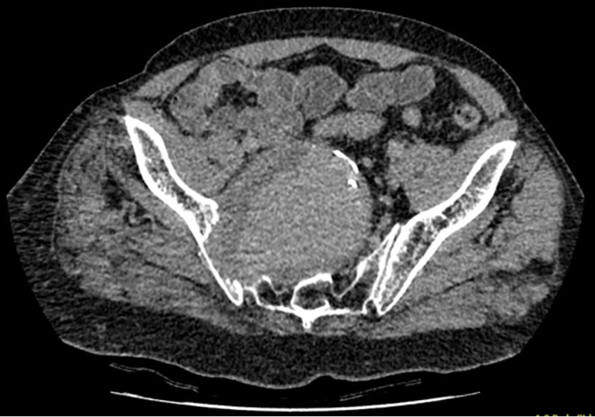
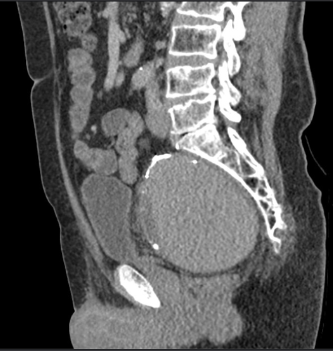
- What do you think could be causing the mass effect?
- A CT scan was requested to further evaluate this patient. See images below. What do you notice?
Click here for answers.
Conclusions
The patient was referred urgently to the vascular surgeons and underwent successful surgery. His hip pain completely disappeared!